
Software for veterinary diagnostics
Acquisition, processing, and management of radiographic images with tools designed for veterinary medicine.
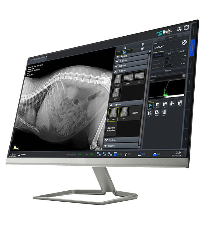
Software is at the heart of every digital radiology system. That is why IBIS offers solutions designed for veterinary diagnostics, capable of simplifying daily tasks and ensuring high-quality images from the very first acquisition.
An intuitive interface, optimized workflows, and dedicated tools help reduce operating times and improve diagnostic efficiency. Each configuration can be customized to meet the needs of the facility, from a small veterinary practice to specialist clinics.
The right software makes all the difference
Tools dedicated to veterinary medicine
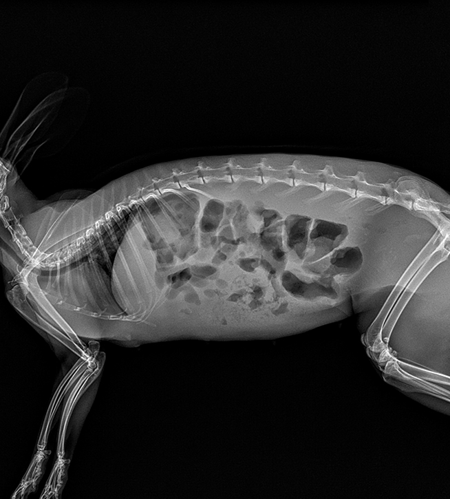
The performance of a radiology system does not depend solely on its hardware. Advanced software enhances every acquisition through sophisticated processing algorithms, automatic image optimization, and tools specific to veterinary anatomy.
Dedicated presets, quick adjustments, and enhancement features deliver detailed images that are immediately ready for diagnostic assessment. The result is a faster, more precise, and more reliable workflow, benefiting both the operator and the patient.
A fast workflow and images always available
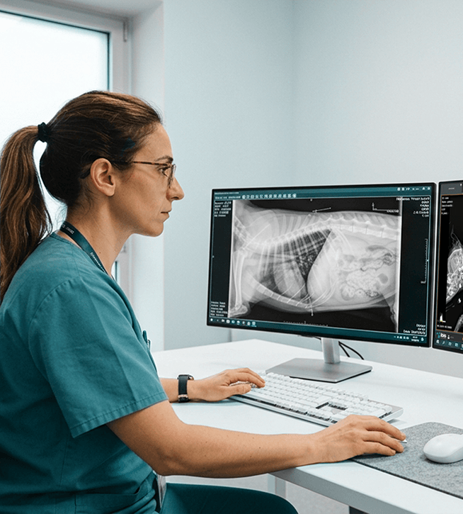
From acquisition to archiving, the software supports every stage of the radiographic examination. Images can be viewed, processed, compared, and shared simply and quickly, providing immediate access to diagnostic information.
Measurement and annotation tools, together with archive management features, make it possible to organize clinical examinations effectively and maintain a complete patient history. Compatibility with DICOM standards and leading PACS systems also ensures seamless integration with the digital infrastructure already in place at the veterinary facility.
Veterinary software
Technical specifications
Veterinary-Specific Measurement Tools
- VHS (Vertebral Heart Score): Automatic calculation of heart size based on the thoracic vertebrae.
- Surgical Planning: Algorithms and step-by-step graphic guides for TPLO and TTA procedures.
- Dysplasia Assessment: Measurement of the Norberg angle and hip distraction index.
- Basic Tools: Geometric measurement of free distances, angles, notes, arrows, and ROI highlights.
Image Processing
- PureImpact™: Proprietary algorithm for optimizing contrast between soft tissues and bone detail.
- Pure Grid: Software suppression of grid lines without using physical grids.
- Auto-Stitching: Automatic stitching of up to 6 images to create full-length spine or long-limb images.
- Auto-Cropping and Auto-Labeling: Cropping, automatic orientation, and insertion of position markers (L/R).
- AI Filters (Noise-X / Bone-X): Advanced deep learning noise reduction and bone suppression for thoracic examinations.
Patient Management and Anatomical Modes
- Species Profiles: Preconfigured menus for dogs, cats, exotic animals, and horses.
- Unlimited APR: Automatic adaptation of exposure parameters to any size or weight.
- Emergency Registration: Start an examination immediately and enter patient details later.
- Multitasking: Management and simultaneous acquisition of examinations for multiple patients.
Connectivity, Standards, and Integrations
- Full DICOM 3.0: Native support for DICOM Storage (PACS), Modality Worklist (MWL), and DICOM Print.
- Hardware Interface: Direct communication with third-party X-ray generators, U-arm systems, collimators, and DAP meters.
- Flexible Export: Save files in native DICOM format or standard JPEG format to USB drives and external disks.
- Touchscreen Interface: Optimized for monitors and tablets, with macro icons, virtual keyboard, and gestures.
- Operating System: Compatible with 64-bit Windows 10 and Windows 11 Pro architectures.
Become our partner
We work with distributors, importers, and hospital groups in major international markets. Request information about our radiology systems, technical documentation, commercial price lists, and dedicated support services.
Contacts
Via Cascina Bruciata, 3, 24068 Seriate – Bergamo
Do you need information for your company?
Contact the IBIS sales office. We respond to inquiries from distributors, importers, hospital groups, and professionals in the medical and radiology sectors.