
Software for Equine Diagnostics
Acquisition, processing and management of radiographic images with tools developed for equine veterinary medicine.
The software offers specific functions for equine radiology, simplifying image acquisition and optimizing the workflow during examinations of the limbs, feet and main equine anatomical structures. Dedicated configurations make it possible to quickly adapt the interface to the operational needs of equine veterinarians, clinics and specialized facilities.
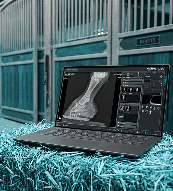
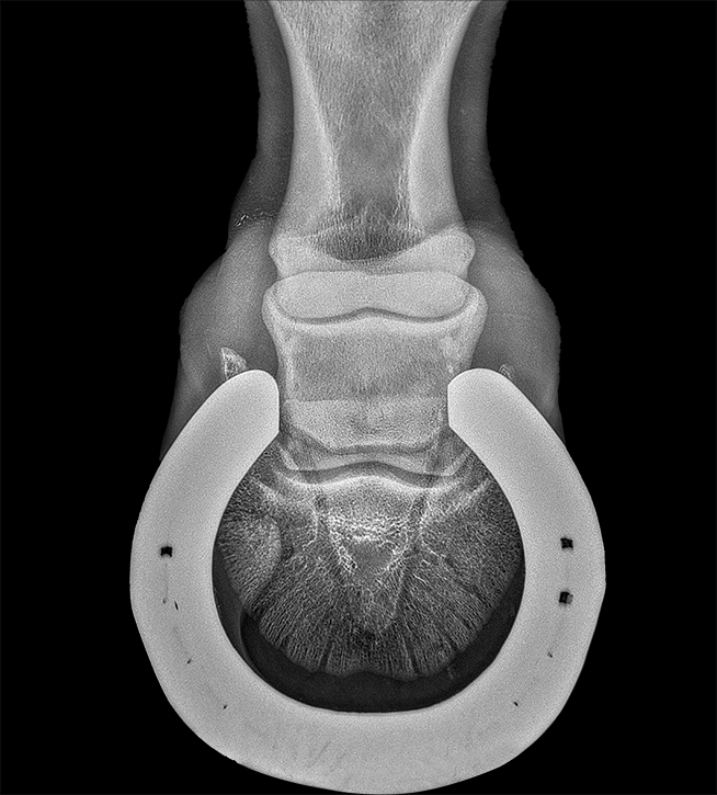
The right software for equine diagnostics
Tools dedicated to equine veterinary medicine
The high number of examinations of the horse’s limbs and extremities calls for fast, accurate and easy-to-use tools. The software integrates dedicated anatomical presets, settings optimized for equine applications and advanced image-processing functions to improve diagnostic quality.
The intuitive interface speeds up daily operations, reduces acquisition times and ensures efficient management of radiographic studies.
A fast workflow for work in the field and in the clinic
From acquisition to archiving, the software supports every stage of the equine radiographic examination. Images can be viewed, processed and shared in seconds, improving collaboration between veterinarians and specialists.
DICOM features enable integration with existing infrastructures, while the examination management tools facilitate the organization of images for sport horses, breeding farms and specialized veterinary facilities.
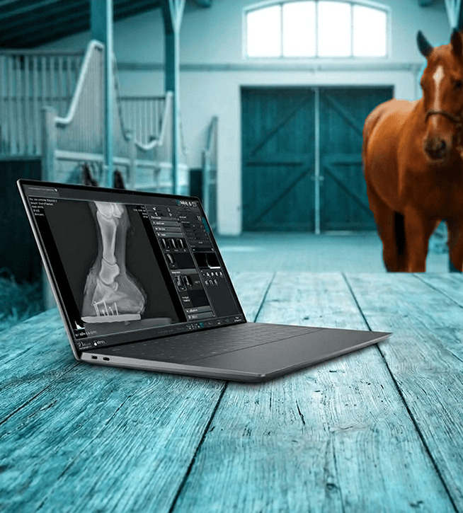
Software for equine veterinary medicine
Technical features
Equine-Specific Measurement Tools
- Advanced Projection Management: Guided selection of standardized projections (e.g. lateromedial, dorsopalmar/plantar) and specific oblique projections for equine orthopedic diagnostics.
- Basic Tools: Geometric measurement of free distances, angles, notes, arrows and ROI highlighting.
- Distal Phalanx Angle (Extensor Process Angle): Measures the inclination of the third phalanx (P3) relative to the ground line to diagnose laminitis or palmar/plantar imbalance.
- Phalanx Alignment Ratio (HPA – Hoof-Pastern Axis): Automatic assessment of the hoof-pastern axis to verify anatomical alignment between the first, second and third phalanges.
- Sole Thickness and Sinking: Millimetric calculation of the distance between the apex of the third phalanx and the hoof sole.
Image Processing
- PureImpact™: Proprietary algorithm for optimizing contrast between soft tissues and bone details.
- Pure Grid: Software-based suppression of grid lines without using physical grids.
- Auto-Stitching: Automatic merging of up to 6 images for entire columns or long limbs.
- Auto-Cropping and Auto-Labeling: Cropping, automatic orientation and insertion of position markers (L/R).
- AI Filters (Noise-X / Bone-X): Advanced deep-learning noise reduction and bone suppression for thoracic examinations.
Patient Management and Anatomical Modes
- Unlimited APR: Automatic adaptation of exposure parameters to any size or weight.
- Emergency Registration: Immediate start of the examination, with patient details entered later.
- Multitasking: Management and simultaneous acquisition of examinations for multiple patients.
- Specific Anatomical Protocols: Schematic graphic maps of the horse’s skeleton for quick projection selection (e.g. fetlock, carpus, hock, stifle, hoof).
Connectivity, Standards and Integrations
- Full DICOM 3.0: Native support for DICOM Storage (PACS), Modality Worklist (MWL) and DICOM Print.
- Hardware Interface: Direct communication with third-party radiographic generators, U-arm systems, collimators and DAP meters.
- Flexible Export: Saving files in native DICOM format or standard JPEG on USB drives and external disks.
- Touchscreen Interface: Optimized for monitors and tablets with macro icons, a virtual keyboard and gestures.
- Operating System: Compatible with 64-bit Windows 10 and Windows 11 Pro architectures.
Become our partner
We work with distributors, importers, and hospital groups in major international markets. Request information about our radiology systems, technical documentation, commercial price lists, and dedicated support services.
Contacts
Via Cascina Bruciata, 3, 24068 Seriate – Bergamo
Do you need information for your company?
Contact the IBIS sales department. We respond to requests from distributors, importers, hospital groups, and professionals in the medical and radiological sector.