
Vivix 1717V Wired X-ray Panel
Reliability and continuity for veterinary radiology
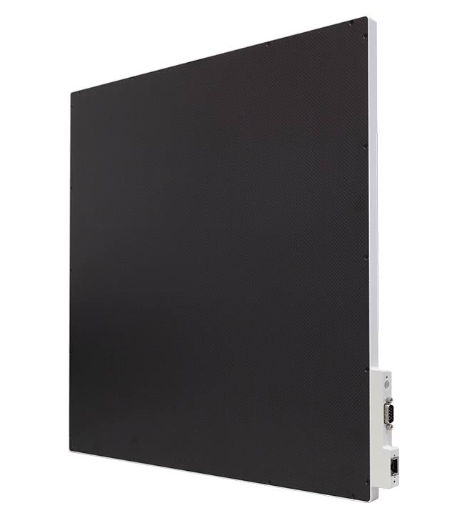
The Vivix 1717V is a 43 × 43 cm digital detector designed for use in fixed veterinary radiology installations.
The large acquisition area allows animals of different sizes to be examined and extensive anatomical regions to be captured in a single exposure.
The wired connection ensures stable communication with the acquisition workstation and keeps the panel always available, without requiring battery management or recharging.
A reliable solution for veterinary practices and clinics looking for operational continuity, ease of use, and integration with existing radiology tables.
Digital Panels
Vivix 1717V
The square 43 × 43 cm format offers a large acquisition area and reduces the need to rotate the panel when switching between different projections.
The digital detector uses a cesium scintillator and is connected directly to the acquisition system via cable. The wired configuration ensures continuous power and stable image transfer, making it particularly suitable for installation inside the cassette holder of a radiology table.
The Vivix 1717V can be supplied as a complete digital system including computer, monitor, and veterinary software, or used to upgrade existing radiology equipment.
The absence of batteries reduces recharging tasks and keeps the panel immediately available throughout the working day.
The VxVue software
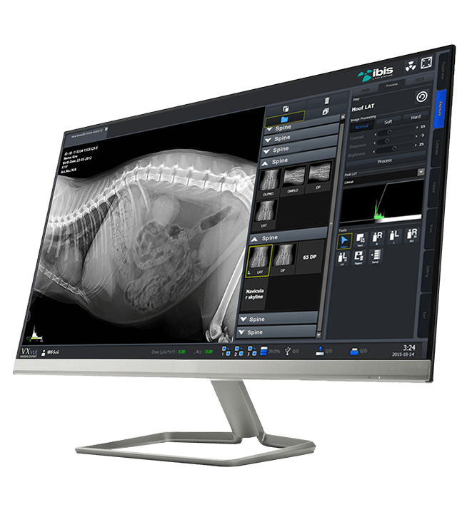
The panel comes with VXvue, the software dedicated to the acquisition, processing and management of veterinary radiographic images.
PureImpact™ algorithms automatically optimize images based on the species and anatomical region, improving detail visibility and tissue definition.
VXvue integrates specific protocols for dogs, cats and exotic animals, as well as guided tools for veterinary measurements: TTA, TPLO, VHS, Norberg angle and Hip Distraction Index. Compatible with the DICOM 3.0 standard, it enables image management, export and transmission to archiving systems.
- Advanced image processing
- Veterinary anatomical protocols
- TTA, TPLO, VHS, Norberg and Distraction Index measurements
- DICOM 3.0 compatibility
Vivix 1717V
Veterinary applications
Orthopedic examinations
- Forelimbs and hindlimbs
- Pelvis and hips
- Spine
- Pre- and post-operative checks
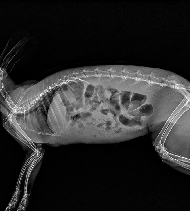
Thoracic and abdominal examinations
- Chest
- Abdomen
- Respiratory system
- Gastrointestinal system
Professional settings
- Veterinary practices
- Veterinary clinics and hospitals
- Examinations of dogs, cats, and exotic animals
- Home radiography
Vivix 1717V
Technical specifications
- a-Si TFT PIN Diode Cesium (CsI) technology
- Wired connection
- Power supplied via the included I/O box
- Pixel Pitch 140 microns
- Format 43 x 43 cm (17″x17″)
- Resolution 3.5 line pairs per millimeter (3.5 lp/mm)
- 3072 x 3072 pixels
- Image size 43 x 43 cm
- 16-bit grayscale
- DQE 57 @ 0.5 lp/mm – MTF 90 @ 0.5 lp/mm
- Gigabit Ethernet interface (1000BASE-T)
- AED/DR Trigger Mode generator interface
- Dimensions 46 x 46 x 1.55 cm
- Weight 4.6 kg
Contacts
Via Cascina Bruciata, 3, 24068 Seriate – Bergamo
Do you need information for your company?
Contact the IBIS sales office. We respond to inquiries from distributors, importers, hospital groups, and professionals in the medical and radiology sectors.