
Vivix 3643FAW Wireless Radiography Panel
Versatility and detail in equine diagnostics
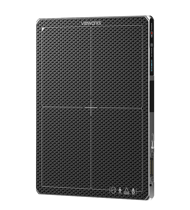
The Vivix 3643FAW combines the large 36 × 43 cm format with the high-resolution Glass-Free technology of the F series.
The acquisition surface allows different anatomical areas to be examined while remaining easy to handle during use in clinics, stables, or equestrian centers.
The 99 µm pixel pitch delivers highly detailed images, while the structure without traditional glass support increases the detector’s durability in everyday use.
A versatile solution for the equine veterinarian who needs a single panel to perform a wide variety of orthopedic examinations.
Digital Panels
Vivix 3643FAW
The 36 × 43 cm acquisition area provides coverage suitable for examinations of the limbs and larger joints.
The Glass-Free a-Si TFT sensor uses a flexible cesium scintillator, 99 µm pixels, and a 3548 × 4316 pixel matrix.
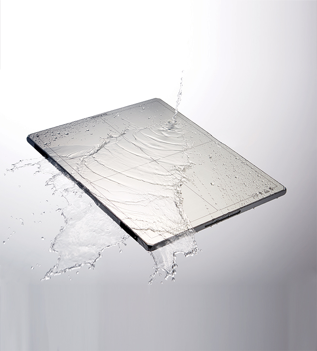
The robust design is tested to withstand drops from a height of up to 1.5 meters, while the IP67 protection shields the device from dust and liquids.
The two batteries provide a stated battery life of up to 16 hours on standby, corresponding to approximately 3,000 images with a 15-second cycle.
Wireless communication makes it easier to use around the horse, limiting cables in the working area.
VXvue software specifically designed for equine veterinary medicine
The panel comes with VXvue, the software dedicated to the acquisition, processing and management of radiographic images.
Advanced PureImpact™ post-processing algorithms optimize images by enhancing the visibility of anatomical details, tissue definition and the overall quality of the examination.
The equine veterinary medicine section includes anatomical protocols specifically designed for horses, with projections and parameters that are preset and customizable by the operator. The software also enables the rapid management of pre-purchase radiographic examinations, organizing the projections to be performed in sequence, and provides guided tools for specialized measurements in equine medicine.
Compatible with the DICOM 3.0 standard, VXvue allows images to be managed, exported and sent to archiving systems.
- Advanced image processing
- Anatomical protocols specifically designed for equine veterinary medicine
- Management of pre-purchase examinations
- Guided specialized measurements
- DICOM 3.0 compatibility
Vivix 3643FAW
Applications in equine veterinary medicine
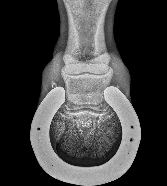
Distal limb examinations
- Foot and hoof
- Phalanges
- Fetlock
- Pastern
Distal limbs
- Carpus
- Tarsus
- Stifle
Professional activities
- Clinic visits
- Examinations at the stable
- Radiographs in the field
- Pre-purchase evaluations
Vivix 3643FAW
Technical specifications
- a-Si TFT technology – Cesium (CsI)
- Pixel pitch 99 microns
- Format 36 x 43 cm (14″x17″)
- Resolution 4 line pairs per millimeter (4lp/mm)
- 3548 x 4316 pixels
- Image size 36 x 43 cm
- 16-bit grayscale
- DQE 57 @ 0,5 lp/mm – MTF 90 @ 0,5 lp/mm
- Multi-Frame Mode 6 fps (10 s per full image)
- Gigabit Ethernet IEEE 802.11n/ac interface
- Generator interface AED/DR Trigger Mode
- IP67 protection rating
- Weight 2.6 kg including 2 batteries
- Dimensions 38.4 x 46 x 1.5 cm
Contacts
Via Cascina Bruciata, 3, 24068 Seriate – Bergamo
Do you need information for your company?
Contact the IBIS sales office. We respond to inquiries from distributors, importers, hospital groups, and professionals in the medical and radiology sectors.