
CDR digital vet
Direct Digital Radiography for Veterinary Medicine
Designed to meet the needs of modern veterinary facilities, the CDR digital vet system enables high-quality radiographic images to be acquired quickly and efficiently. Direct digital technology eliminates development times, streamlines the workflow, and makes images immediately available for diagnostic evaluation.
Thanks to its high image quality and dedicated software tools, the system supports veterinarians in their daily diagnostic activities, helping to improve the facility’s operational efficiency and the animals’ comfort during examinations.
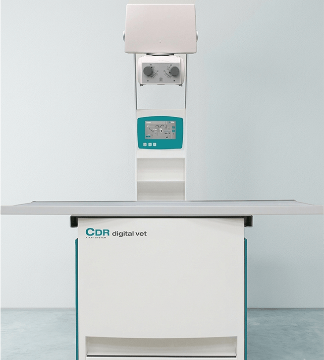
CDR digital vet
Clinical applications
- Small animal diagnostics
- Ideal for radiographic examinations of dogs, cats, and companion animals, with images available in seconds for immediate evaluation.
- Veterinary orthopedics
- Particularly suitable for evaluating fractures and joint disorders, pre- and post-surgical checks, and monitoring orthopedic treatments.
- Thoracic and abdominal diagnostics
- Enables radiographic imaging of the chest and abdomen to support diagnostic and therapeutic activities.
- Pre- and post-surgical examinations
- Supports surgical planning and clinical monitoring activities thanks to the immediate availability of digital images.
- Veterinary emergency care
- The system’s immediate availability makes it possible to quickly perform radiographic examinations on patients requiring urgent evaluation.
CDR digital vet
Advantages
- Integrated direct digital detector
- The DR system enables high-definition radiographic images to be acquired and viewed in seconds, improving diagnostic speed and reducing waiting times.
- Fully digital workflow
- From image acquisition to viewing, every stage of the examination is managed digitally, streamlining the workflow and increasing the veterinary facility’s productivity.
- Complete radiology solution
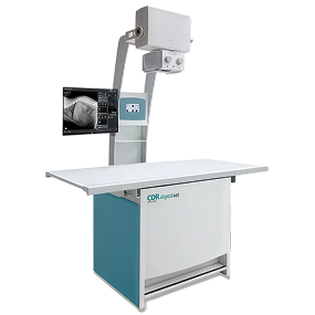
- CDR digital vet integrates a radiographic table, generator, and DR acquisition system into a single workstation designed to ensure ease of operation and high performance.
CDR digital vet
Technical Specifications
Technical specifications
Technology:
Digital
Acquisition, post-processing, archiving, and export software
Connection to the hospital network
DICOM compatibility
RIS / PACS interface
Worklist acquisition
Generator:
32kW, 125kV, 425mA, 100kHz high-frequency generator
IBIS HF1 100/2 Inverter model
kV range 40-125 kV in 1kV steps
mAs range 0.2-220 mAs in 28 steps
2- and 3-point radiographic techniques
Can be limited to 99kV if necessary
Acquisition system:
Direct digital detector, 43x43cm (17″x17″) format
Desktop workstation with an additional disk for study archiving
Collimator:
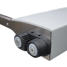
Square field 0x0cm – 43x43cm (SID 100cm)
LED lamp
30-second ON/OFF timer
Guide for inserting accessories
Retractable measuring tape
Operational features:
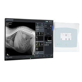
Color touchscreen display for controlling radiographic parameters
External monitor for viewing the acquisition software interface
Option: column-mounted monitor with articulated arm and internal fanless computer
Customizable Anatomically Programmed Radiography (APR)
Interconnection between acquisition software and X-ray generator
Four-way floating tabletop
Soundproof mechanical brakes
Antibacterial tabletop
Automatic shutdown after 30 minutes
Safety pedal for X-ray exposure
Dimensions:
Patient table measuring 1,500 mm x 745 mm
Optional patient table measuring 1,800 mm x 745 mm
Patient table height from floor: 775 mm
Maximum overall dimensions (standard tabletop version): 2,050 mm x 1,128 mm
Maximum overall dimensions (extended tabletop version): 2,350 mm x 1,128 mm
Power supply:
Single-phase 230 Vac +/-10% 50/60Hz
Power consumption: 0.5 A standby; 230 Vac – 12 A pulsed
Become our partner
We work with distributors, importers, and hospital groups in major international markets. Request information about our radiology systems, technical documentation, commercial price lists, and dedicated support services.
Gallery
Contacts
Via Cascina Bruciata, 3, 24068 Seriate – Bergamo
Do you need information for your company?
Contact the IBIS sales department. We respond to requests from distributors, importers, hospital groups, and professionals in the medical and radiological sector.